On study days 0 and 7, mice are anesthetized with Isoflurane and sensitized with a 0.1 mL intradermal injection at the base of the tail consisting of an equal mixture of 4 mg/mL Mycobacterium tuberculosis (MTB) in Freund’s complete adjuvant (CFA) and 4 mg/mL methylated bovine serum albumin (mBSA) in water. On study day 10, mice are injected into the right hind footpad with 20 µL of 10 mg/mL mBSA.
Disease Parameters:
The Type IV DTH reaction is an acute inflammation driven by cell-mediated immunity.1 Typically, a DTH response develops as a result of a 1 to 2 week sensitization period following primary antigen contact in which dendritic cells and macrophages interact with an antigen and present to T cells in the regional lymph nodes. During this time TH cell recruitment, activation and clonal expansion of predominantly CD4+ Th1 cells occurs. Collectively, this process as known as the Sensitization Phase.2 A follow-on exposure to the antigen initiates the Effector Phase of the DTH response, in which Th1 cells secrete a variety of cytokines that recruit and activate macrophages and other non-specific inflammatory cells. This results in a visible inflammatory response within 24 to 48 hours post antigen challenge. The delayed response is a direct result of the time needed for cytokine secretion and recruitment/activation of macrophages. More specifically, the DTH response is mediated by T helper cells secreting predominantly interferon-g and IL-2 with production of inflammatory mediators and subsequent leukocyte recruitment. The mechanism by which leukocytes are attracted to the site of antigen re-introduction is not clear, but multiple cytokines and chemokines are thought to be involved in the DTH reaction.
Dosing Paradigms:
Begin dosing on study day 14, 0, or 8 and continue until necropsy on day 11 (24 hours after mBSA challenge).
Route of administration: SC, PO, IP, IV, topical
Clinical Assessment:
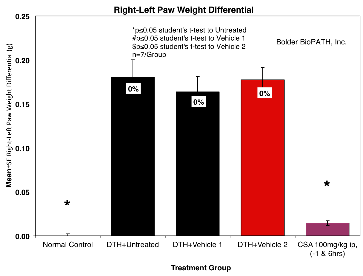
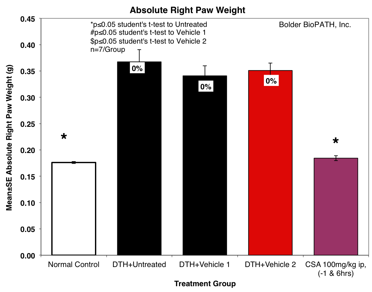
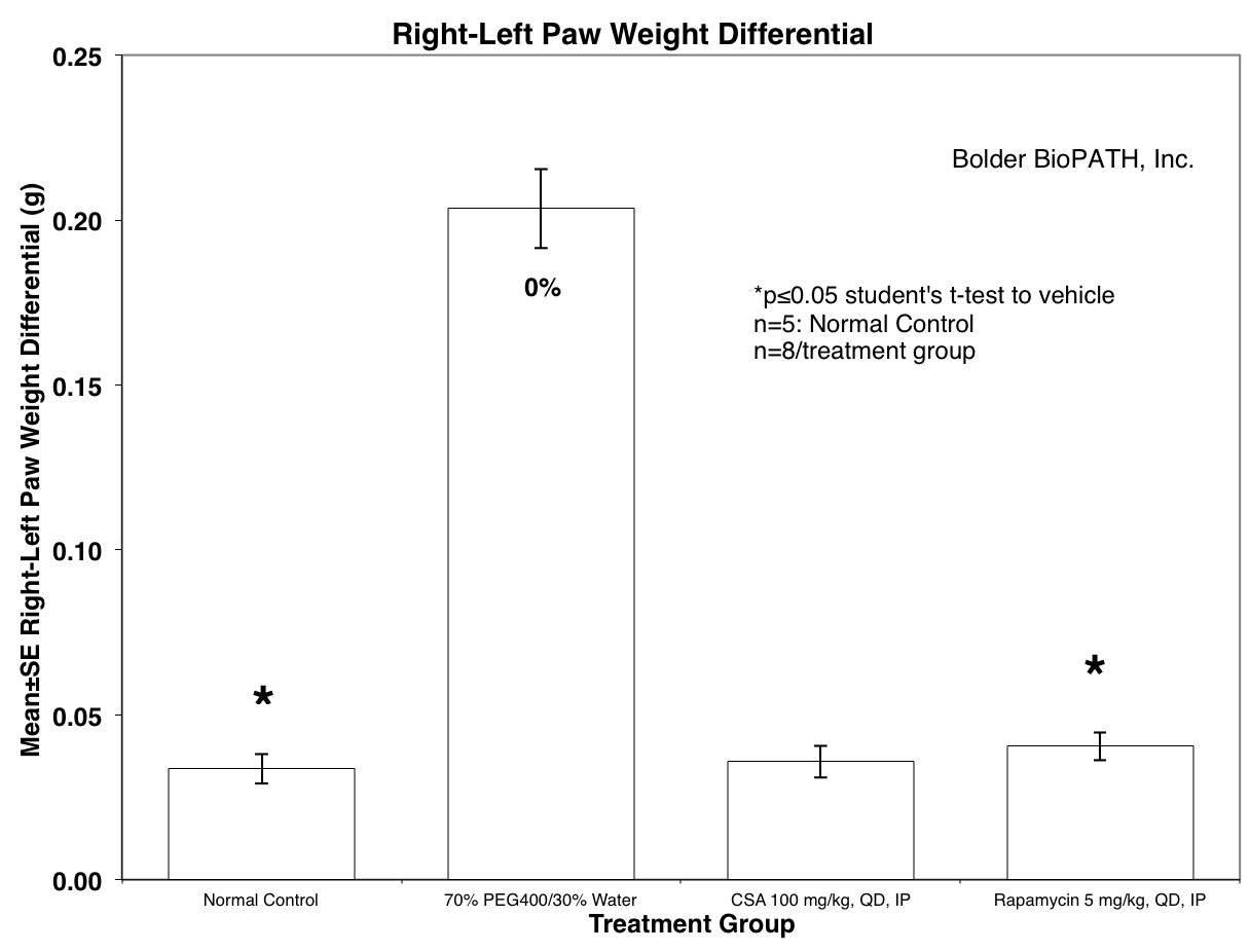
Measurement of the DTH reaction can be done grossly by comparing the paw weight difference between normal versus antigen-injected paws. Right and left hind paws are collected, transected at the medial and lateral malleolus, and weighed.
Sample Data:
For examples of positive controls, please contact us.
Notes:
Chemokines such as IL-8, monocyte chemoattractant protein 1 (MCP-1), macrophage inflammatory protein 1a (MIP-1a), and macrophage migration inhibitory factor (MIF) have been found to be involved in the recruitment of leukocytes to the DTH reaction site. Cytokine inhibitors, such as anti-IL-16, have been shown to minimize the DTH response. Other compounds that effectively inhibit the DTH reaction are dexamethasone (a potent steroid that induces lympholysis) and cyclosporine-A (CsA), which inhibit the action and growth of T-lymphocytes.
Optional Endpoint:
PK/PD blood collections
Cytokine/chemokine analysis via Luminex(R)
Other sandwich ELISAs
CBC/clinical chemistry analysis
Soft tissue collection
Histopathologic analysis
Immunohistochemistry analysis
References:
Yoshimoto T, Wang CR, Yoneto T, et al. Role of IL-16 in delayed-type hypersensitivity reaction. Blood. 2000;95(9):2869–2874.
Owen J, Punt, J, Stranford S. Kuby Immunology. 7th ed. New York: WH Freeman & Company; 2013. Chapter 15, Inflammation: Allergy and Hypersensitivities.